Figure 3.
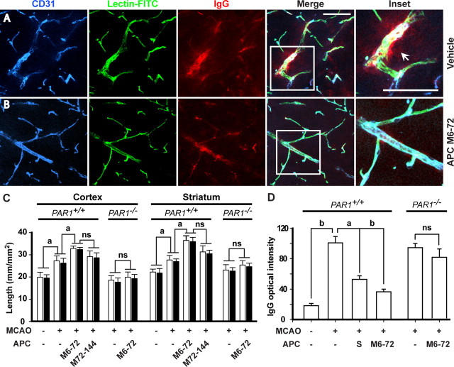
APC therapies at 6–72 and 72–144 h after transient ischemia enhance neovascularization in the ischemic border in mice via PAR1. A, B, Expression of CD31 (brain endothelial marker), in vivo systemic lectin-FITC angiography, and serum IgG leakage studied simultaneously by three-color confocal laser-scanning microscopy in the ischemic border in the striatum 7 d after 1 h transient MCAO in vehicle-treated mice (A) or APC-treated mice (APC at 0.8 mg/kg, i.p., at 6, 24, 48, and 72 h after ischemia) (B). Inset was taken from the merged image. Scale bar, 50 μm. CD31, blue; lectin-FITC, green; IgG, red. C, Total capillary length of CD31-positive structures (open bars) and of in vivo lectin-perfused vessels (closed bars) in the cortex and striatum in the periinfarct regions 7 d after 1 h transient MCAO in mice treated with vehicle and APC multiple doses M6–72 (0.8 mg/kg, i.p., at 6, 24, 48, and 72 h post-MCAO) and M72–144 (0.8 mg/kg, i.p., at 72, 96, 120, and 144 h post-MCAO), and in PAR1-null mice (PAR1−/−) treated with vehicle or APC multiple-dose M6–72. D, Quantification of the IgG signal intensity in the striatum 7 d after 1 h transient MCAO in mice treated with vehicle, APC multiple-dose M6–72, or APC single dose (1.6 mg/kg, i.p.; S) 24 h post-MCAO, compared with sham-operated controls and in PAR1−/− mice treated with vehicle or APC multiple-dose M6–72. Shown are mean ± SEM (n = 5–6 mice per group). For sham-operated control mice, n = 3. ap < 0.5; bp < 0.01; ns, nonsignificant.

