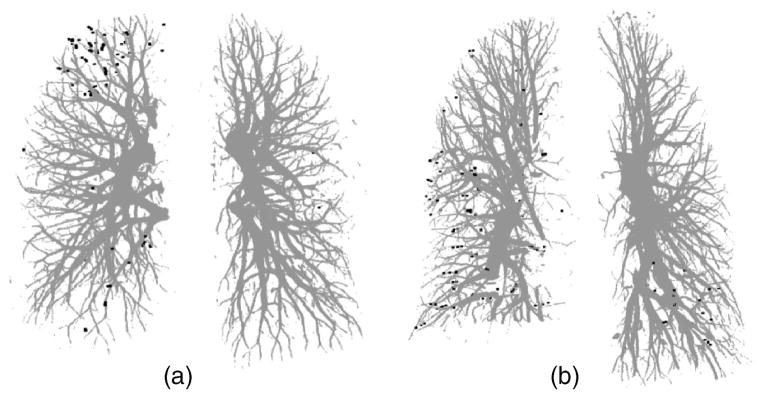
Fig. 8.
False negative vessel center points indicating missed vessels by our automated vessel segmentation method compared to radiologist’s manual tracking for (a) the test case without other disease and (b) the test case with pleural effusion disease. All of the false negative vessel center points were located in subsegmental peripheral vessels for both test cases.

