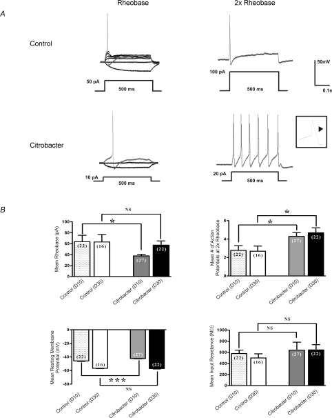
Figure 1. Effects of C. rodentium induced colitis on excitability of colonic DRG.
neurons A, representative current clamp trace of action potential elicited at rheobase and two times rheobase from labelled neurons isolated from C. rodentium treated and control animals. The amount of current needed to elicit an action potential is significantly lower in labelled C. rodentium neurons (10 pA) compared to control neurons (50 pA). Furthermore, the C. rodentium neurons fire significantly more action potentials at 2× rheobase compared to control neurons. Action potential tracings from control neurons are shown in upper panel and those from C. rodentium treated neurons are shown in lower panel. A 500 ms depolarising current pulse was used to elicit the action potentials. Inset shows one of the action potentials using expanded time scale to exhibit the characteristic hump on the falling phase typical of nociceptive neurons. B, data summarizing changes in electrophysiological properties of nociceptive DRG neurons during acute infection (day 10) and following resolution of infection (day 30). C. rodentium induced colitis caused a significant reduction in rheobase, significant increase in the mean number of action potentials at 2× rheobase and a significant reduction in the mean resting membrane potential in labelled neurons isolated from C. rodentium treated (grey filled bars) animals during acute infection compared to matched controls (stippled bars). The mean input resistance did not change significantly. Data are means ±s.e.m. *P < 0.05; NS, not significant. At day 30, neurons from C. rodentium treated (black filled bars) animals fired significantly more action potentials at 2× rheobase compared to controls (open bars) whereas, the mean rheobase, resting membrane potential and input resistance returned to comparable control values. Data are means ±s.e.m. *P < 0.05.

