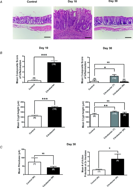
Figure 3. Sustained hyperexcitability of colonic DRG neurons following resolution of C. rodentium induced colitis.
A, representative photomicrographs of H&E staining of colonic tissue sections illustrating normal histology of control tissues, increased inflammation and crypt height during active infection (day 10) and return to near normal colonic mucosal histology on resolution of infection (day 30). Scale bar = 100 μm. B, microscopic histological colitis scores determined by a pathologist blinded to the treatments. Mean composite scores of inflammation are shown in upper panels and mean crypt heights are shown in the lower panels for active infection on day 10 (left panels) and resolved infection on day 30 (right panels). For day 30 colons, T (all colons) and NI (non-inflamed colons only) based on microscopic inflammation scores. Values show a marked increase in inflammation at day 10 which had resolved in most colons by day 30 although some still showed subtle evidence of inflammation. ***P < 0.0001, **P < 0.005. C, summary data illustrating persistent hyperexcitability of nociceptive DRG neurons following resolution of infection (day 30). Neurons from C. rodentium treated animals fired significantly more action potentials at 2× rheobase compared to control. *P < 0.05. The mean rheobase was lower, but not significant in the C. rodentium treated neurons compared to controls.

