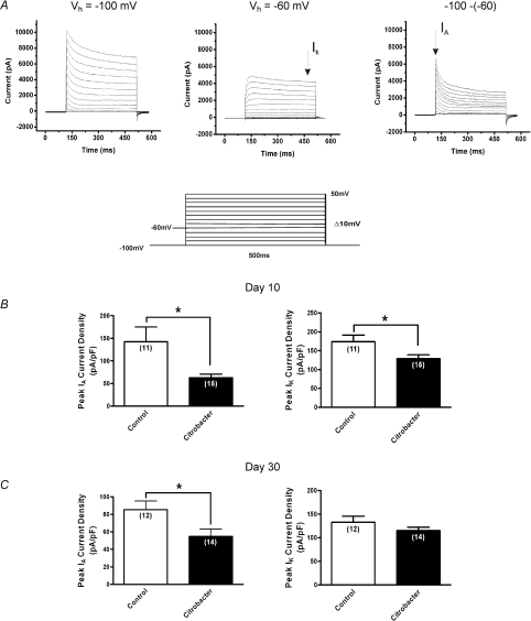
Figure 5. Effects of C. rodentium induced colitis on isolated voltage gated Kv currents.
A, representative voltage clamp traces showing outward voltage gated K+ current families separated biophysically by manipulating the holding potential. Currents were generated using a 10 mV voltage step protocol (illustrated in inset) from −90 mV to +50 mV. At holding potential of −100 mV (left), two currents were evident, a transient inactivating A-type current (IA) and sustained non-inactivating IK type currents. At a holding membrane potential of −60 mV, IA current was significantly inactivated such that only the sustained component IK is elicited (middle) and subtraction of the sustained from the total current yielded IA (right). B, summary of peak current density for IA and IK respectively. Current densities were obtained by normalizing the measured amplitudes of the peak transient components of the isolated currents to the cell capacitance. At the height of inflammation (day 10), C. rodentium induced colitis caused significant suppression of both IA and IK current densities (filled bar) compared to controls (open bar). C, following resolution of infection (day 30), the reduction in IA current density was sustained in neurons from C. rodentium infected animals (filled bar) compared to controls (open bar). There was no significant difference in IK current density between the two groups of neurons. Data are means ±s.e.m. *P < 0.05.

