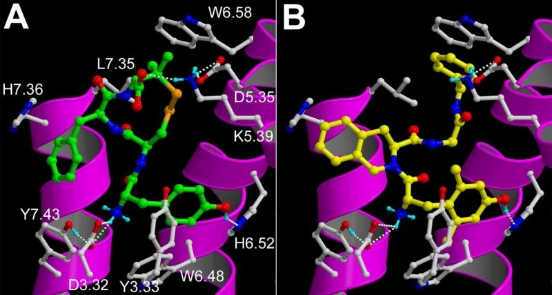
Figure 3.
Docking poses of: (A) JOM13 (green carbon atoms) and (B) compound 4 (yellow carbon atoms) in the DOR receptor model. The backbone of transmembrane helices 5, 6, and 7 are represented by magenta ribbons (TM3 is not shown for clarity). Important binding residues are depicted as ball-and-sticks with grey carbon atoms. Oxygen, nitrogen, sulphur and hydrogen atoms are coloured red, blue, orange and cyan, respectively. H-bonds described in the text are depicted by white dots.

