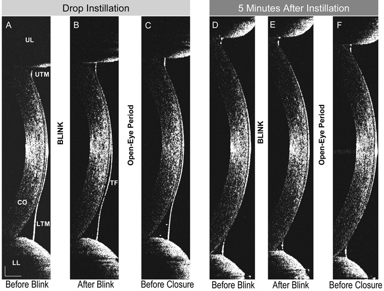
Figure 1. OCT images obtained during blinking.
A vertical 12 mm OCT scan was performed across the corneal apex during blinking immediately after instillation of artificial tears (Images A–C) and 5 minutes later (Images D–F). Changes in tear film (TF), upper tear meniscus (UTM) and lower tear meniscus (LTM) were noted after blinking and open-eye period. Cornea (CO), upper eyelid (UL) and lower eyelid (LL) are also shown. Bars denote to 500 µm.

