Fig. 1.
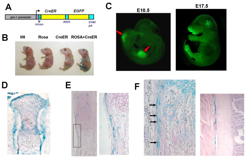
(A) Schematic representation of the Prx1CreER-GFP transgene. cDNAs for CreER and EGFP were cloned downstream of a 2.4 kb Prx1 promoter. (B) X-gal staining of E18.5 embryos showing clear correlation with the genotype. Tamoxifen was injected at E15.5 and E16.5. Only the embryo harboring the Prx1CreER-GFP transgene and Rosa26 LacZ reporter shows positive staining in the limbs. Other embryos show minimal background. (C) GFP fluorescence of Prx1CreER-GFP embryos at E10.5 (left) and E17.5 (right). E10.5 embryo shows fluorescence in the forelimb and hindlimb buds (arrows). E17.5 embryo shows fluorescence in the limbs and some craniofacial regions. (D) X-gal staining of the proximal tibia of E17.0 Rosa26 LacZ; Prx1CreER-GFP embryo. Tamoxifen was injected at E9.0. (E) X-gal staining of the ulna of E18.5 Rosa26 LacZ; Prx1CreER-GFP embryo. Tamoxifen was injected at E15.5 and E16.5. The right panel shows higher magnification of the boxed area in the left panel. (F) X-gal staining of the radius (left) and tibia (right) of a Rosa26 LacZ; Prx1CreER-GFP mouse at P26. Tamoxifen was injected daily between P19–P23. Sections were counterstained with orcein (right) or hematoxylin, eosin, and alcian blue (left). In the radius, cells in the inner layer of the periosteum stain positive for X-gal (arrows). In the tibia, X-gal-stained periosteal cells are closely associated with the cortical bone.

