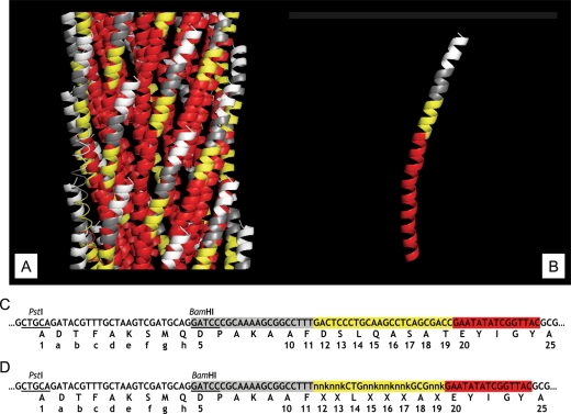
Fig. 1.
Fragment of three-dimensional structure of landscape phage, pVIII protein, fragment of nucleotide sequence of pVIII gene and amino acid sequence of pVIII protein in landscape phages (A). Fragment of three-dimensional structure of landscape phage composed from subunits of major coat protein pVIII (B). White areas correspond to the position of the guest peptides (domain A), grey—to the 5–11 amino acids (domain B), yellow—to the area of 12–19 amino acids ( domain C), and red—to the area of 20–24 amino acids exposed at the phage surface ( domain D). Area corresponding to 25–50 amino acids buried inside the capsid and is not visible. (B) Fragment of three-dimensional structure of pVIII coat protein bearing the β-galactosidase binding peptide. (C) Fragment of DNA and amino acid sequence of mature pVIII in landscape phage 1G40. (D) Fragment of DNA and amino acid sequence of mature pVIII in G-α-library. Numbers correspond to the numbers of amino acids in wild-type pVIII protein.

