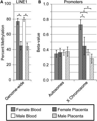
Figure 1.
Reduced placental methylation found at LINE1 repetitive elements and promoters on the X chromosome. Average level of methylation for female blood (grey), female placenta (grey hatched), male blood (white) and male placenta (white hatched) are shown with error bars (one standard deviation) based on the average sample deviation at a single site. Significance calculated using Mann–Whitney test with P < 0.001 (*). (A) LINE1 percent methylation as determined by pyrosequencing at LINE1 repetitive elements across the genome. (B) Illumina Golden Gate Promoter methylation array data averaged separately for 1421 sites on the autosomes and 84 X-linked sites. Beta-values represent average percent methylation.

