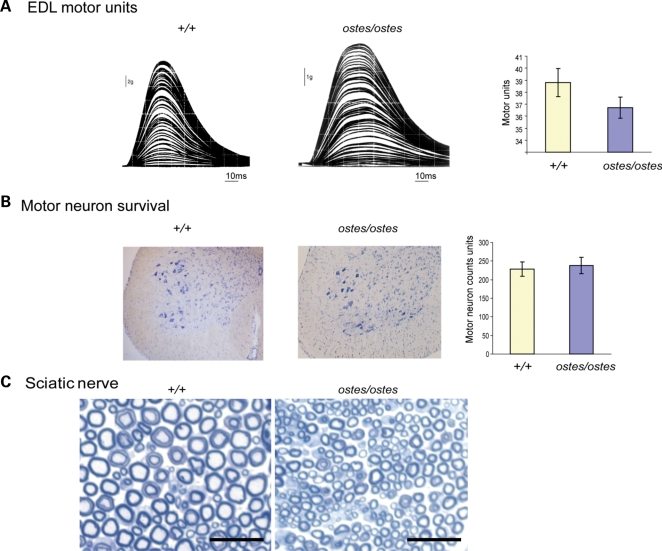
Figure 3.
Motor neuron survival in ostes. (A) Assessment of motor unit number in ostes/ostes EDL. Examples of stepwise increments in twitch force from wild-type (+/+) and ostes/ostes EDL in response to gradual intensity increases of stimulation of the motor nerve to the EDL. Motor unit number in ostes/ostes EDL is not significantly different to wild-type EDL (n = 6 per genotype). Data shown as mean motor unit number ± SEM. (B) Motor neuron survival in ostes/ostes mice. Shown are representative 20 μm-thick transverse sections of spinal cord from wild-type and ostes/ostes mice. Motor neurons are stained with gallocyanin (blue). Data shown as mean motor neuron counts ± SEM. The number of gallocyanin-stained motor neurons present in the sciatic motor pool of the ventral horn of each spinal cord was determined by counting every third serial section (n = 60 sections per mouse; n = 6 per genotype). (C) Representative cross-section of proximal regions of sciatic nerve from 6-week-old wild-type (+/+) (n = 3) and ostes/ostes mice (n = 3) fixed with osmium tetroxide and stained with methylene blue. No demyelination of ostes/ostes motor neurons as evidenced by reduced myelin-to-lumen staining or by ‘onion-bulb’ morphology is seen. Note that ostes/ostes motor neurons are markedly smaller than wild-type, reflecting the overall small size of ostes/ostes mice. Scale bar: 25 µm.

