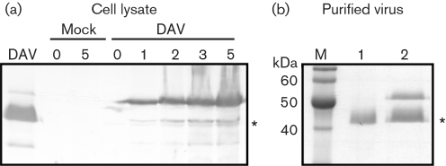
Fig. 2.
Detection of virus protein synthesis in infected Drosophila DL2 cells. (a) DL2 cells were either mock-infected or infected with DAV and incubated at 26 °C until harvesting. Cell samples were taken at 0, 1, 2, 3 and 5 days p.i. and lysed with sample buffer. Cellular proteins were separated by electrophoresis through an SDS–10 % polyacrylamide gel, transferred to a nitrocellulose membrane and incubated with an anti-DAV rabbit polyclonal antiserum to detect virus proteins. The asterisk indicates the 42 kDa capsid protein. Virus proteins were detected at 1 day p.i. and their levels increased over time. No DAV-specific bands were detected in the mock-infected samples. (b) Proteins from DAVHD virions purified from infected DL2 cells (lane 2) and adult Drosophila (lane 1) were electrophoresed through an SDS–10 % polyacrylamide gel stained with Coomassie blue. The asterisk indicates the 42 kDa capsid protein.

