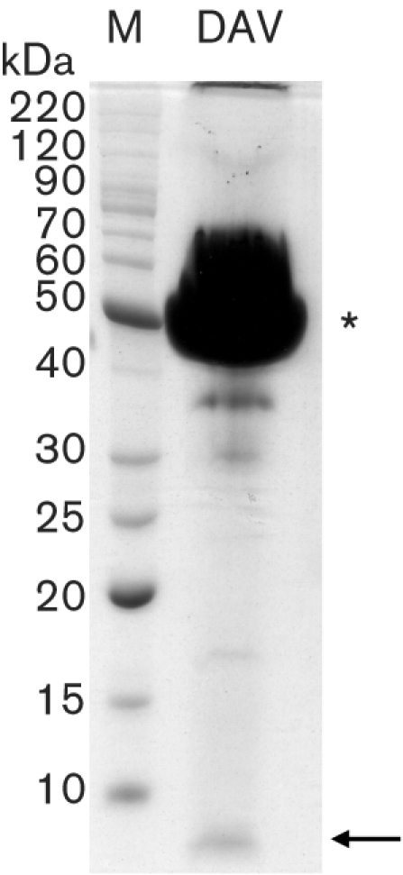
Fig. 3.

Identification of a small structural protein. Virion proteins were overloaded and separated by electrophoresis through an SDS–10 % polyacrylamide gel. Total protein was stained with Coomassie blue. The asterisk indicates the 42 kDa capsid protein band that is consistently observed in SDS-PAGE analyses. The arrow indicates the small protein band that is estimated to be approximately 6 kDa.
