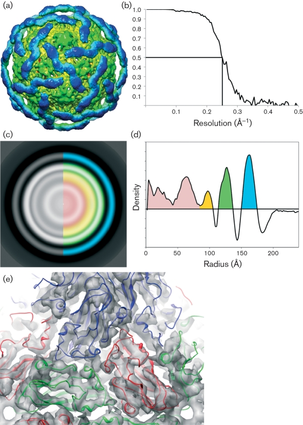
Fig. 7.
Analysis of three-dimensional reconstruction of DAV particles. (a) Surface representation of the DaV cryoEM reconstruction contoured at 2σ. The inner N-terminal domain (green) is well-ordered with visible secondary-structural features, whereas the outer C-terminal domain (blue) contains only very-low-resolution features. (b) Estimated resolution of the reconstruction is 8 Å (0.8 nm) according to FSC criteria (y-axis) at 0.5. (c) Radially averaged slice through the centre of reconstruction. Blue, C-terminal domain; green, N-terminal domain; orange and red, RNA. (d) Plot showing density by radius [colours as in (c)]. (e) Docking of the β-sandwich domain of the TBSV crystal structure into the N-terminal domain of the DaV cryoEM density.

