Fig. 1.
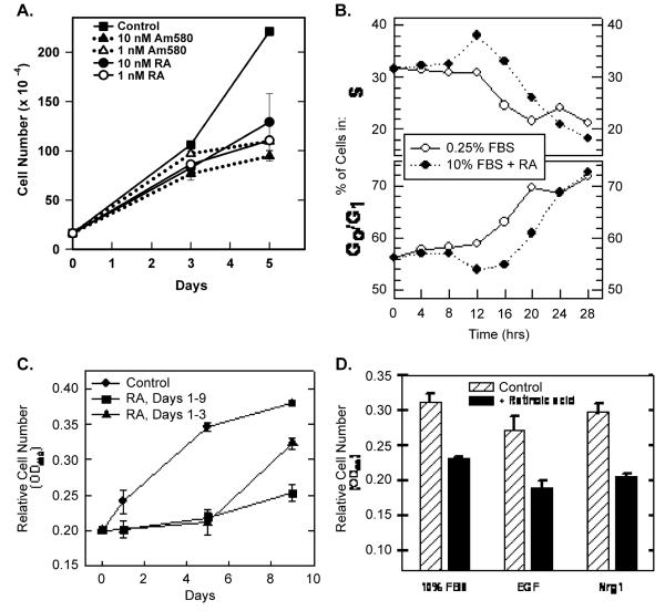
Retinoids reversibly inhibit T-47D cell proliferation. (A) T-47D cells (initial plating density = 1.5 × 10⁵ cells per 60-mm dish) were cultured in the presence of the indicated concentration of RA or the RARα-selective retinoid, Am580. Day 0 represents the time of retinoid addition. Cells were trypsinized and cell numbers determined 3 and 5 days later (data plotted ± SEM). Viability, as determined by trypan blue exclusion, was >90% at all times. (B) The effect of RA and serum deprivation on T-47D cell cycle distribution was determined at 4-h intervals by FACS analysis of propidium iodide-stained cells. The data show the change in the percentage of cells in either S (top) or G0/G1 (bottom). Serum deprivation (after 12 h) and RA treatment (after 16 h, 10−7 M) prevented entry into S. (C) T-47D cells were initially plated at 5 × 10³ cells/well in 96-well dishes (six wells per condition). Relative cell numbers were determined using MTS reduction as measured by absorbance at 490 nm (data plotted ± SEM). RA (10−7 M) was added at day 0 and again at day 3 (squares and triangles). On day 5, RA was removed from half of the treated wells (triangles). (D) T-47D cells were plated at 5 × 10³ cells/well in 96-well dishes in RPMI 1640 + 10% FBS. After 24 h, media were replaced with RPMI 1640 supplemented with 10% FBS, 20 ng/ml EGF or 10 ng/ml Nrg 1 in the absence or presence of 10−7 M RA. After 48 h, relative cell numbers were determined using MTS reduction (±SEM).

