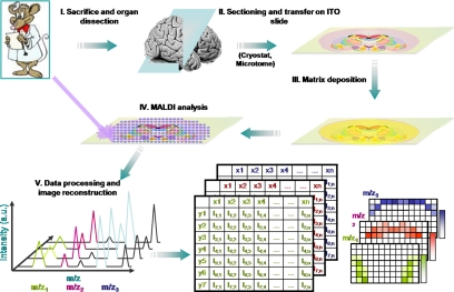
Fig. 1.
Schematic representation of the MALDI-MSI work flow. After tissue sectioning and transfer onto a conductive and transparent sample plate, the MALDI matrix is deposited, and data are acquired by recording mass spectra according to a raster of points covering the surface to be analyzed. Mass spectra recorded with their coordinates on the tissue are processed, and molecular images of the localization of molecules can be reconstructed. a.u., arbitrary units; ITO, idium tin oxide.

