
Figure 3. A Cl channel evolved from a Cl/H exchanger.
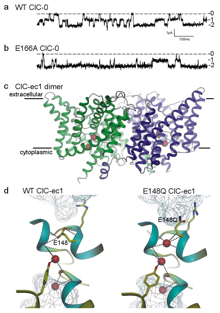
a/, b/ Current recordings showing independent opening and closing of the 2 pores in a wild-type (WT) ClC-0 channel (a), and the mostly open behaviour of an E166A ClC-0 channel (b); labels 0, 1, and 2 indicate number of open pores in each dimeric channel [Modified with permission from ref. 27]. c/ Transmembrane region of WT ClC-ec1 dimer viewed from within the membrane (membrane boundaries indicated). The ion pathway in each monomer (green, blue) contains 2 anions (red spheres). [Modified with permission from ref. 85]. d/ Pore region connecting extra- and intracellular aqueous vestibules (cyan mesh) in WT (left) and E148Q (right) ClC-ec1, containing 2 or 3 Cl ions (red spheres) respectively; the third Cl ion occupies the space vacated by the swung-out side chain of the external gate residue, 148 (E148 is equivalent to E166 of ClC-0). [Modified with permission from ref. 86].