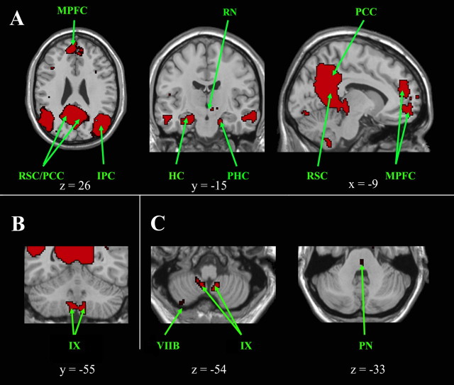
Figure 3.
Cortical, subcortical, and cerebellar regions of the default mode network. A, Cortical and subcortical regions of the default mode network are shown on axial, coronal, and sagittal slices. B, Cerebellar regions are shown on a coronal slice and in C on axial slices, which also highlight a pontine region. The left side of the image corresponds to the right side of the brain. This is an intersection map showing only voxels that were present in the default mode network of both datasets at a corrected threshold of p < 0.01. HC, Hippocampus; IPC, inferior parietal cortex; MPFC, median prefrontal cortex; PCC, posterior cingulate cortex; PHC, parahippocampal cortex; PN, pontine nucleus; RN, red nucleus; RSC, retrosplenial cortex.

