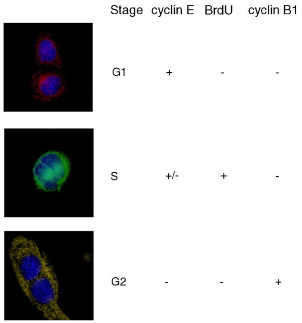
Fig. 2.
Identifying cell cycle populations using immunohistochemical staining. Columns from left to right: Fluorescence image of cell pair, cell-cycle phase assignment, staining for cyclin E, BrdU and cyclin B1. Top row: G1-phase population. Middle row: S-phase population. Bottom row: G2-phase population.

