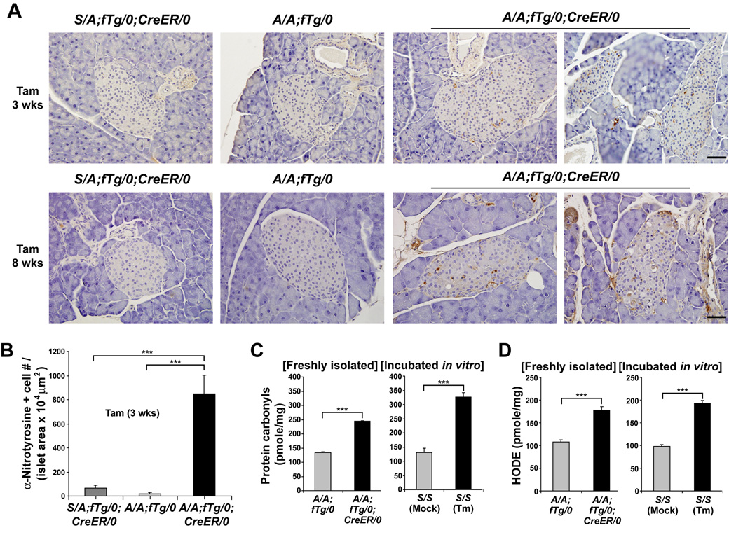
Figure 5. Translation attenuation is required to prevent oxidative stress in beta cells.
(A and B) Immunohistochemistry was performed to detect nitrotyrosine in pancreatic sections from mice at 3 and 8 wks after Tam administration. Representative images and quantitation of the number (#) of nitrotyrosine-positive (+) beta cells per islet area are shown. n = 5–7 mice per group. The scale bars represent 50 µm.
(C and D) Protein carbonyls (C) and HODEs (D) were quantified in extracts from islets freshly isolated from mice at 3 wks after Tam injection. n= 7 mice per group. Islets were isolated from wt S/S mice and in vitro incubated in the absence or presence of tunicamycin (Tm, 2 µg/ml) for 5 hrs prior to analysis of oxidation products. Data are Mean ± SEM, (B–D).

