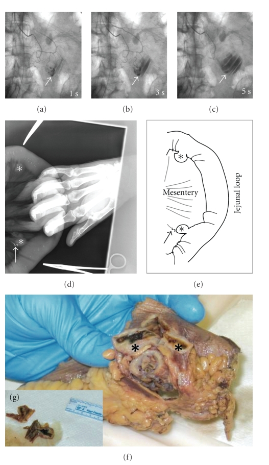
Figure 1.
Massive gastrointestinal bleeding from an ArterioVenous malformation within a jejunal diverticulum. Superior Mesenteric Angiography (a)–(c) revealed active extravasation (arrow) of the contrast material in the proximal jejunum and pooling of the same (arrow) in the jejunum (c). Intraoperative plain film X-ray obtained during segmental resection of the proximal jejunum (d, e) revealed the presence of two diverticula (stars), located on the mesenteric side. The arrow marks the diverticulum that was the source of hemorrhage, as determined by the presence of the radio opaque angiographic coil which was previously deployed during angiographic intervention. The specimen from segmental jejunal resection (f, g) shows blood clot within the diverticula.

