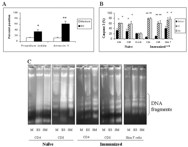
Fig. 1. 1 × 10⁶ cells isolated from the skin or skin-draining lymph nodes of naive or immunized animals were cultured in medium alone (M) or with ES products (60 μg/ml) or 100–150 schistosomula (SM) for 24 h.
Following incubation, apoptosis was measured by annexin V and propidium iodide staining and counting 500 –1000 cells under a fluorescence microscope (A), by measuring caspase-3 activity in various subset of lymphocytes using PhiPhiLux substrate and counting 500 –1000 cells under a fluorescence microscope (B), and by evaluating DNA fragmentation in different subsets after treatment with DNA digestion buffer and separation on a 1% agarose gel (C). Data presented are representative of one of three to five similar experiments using five to seven mice per group in each experiment. A and B show means ± S.D. of the percentage of positive cells. * and **, p < 0.05 and p < 0.01 compared with the medium control, respectively.

