
Fig. 2. Time courses of caspase-8 and caspase-3 protease activities.
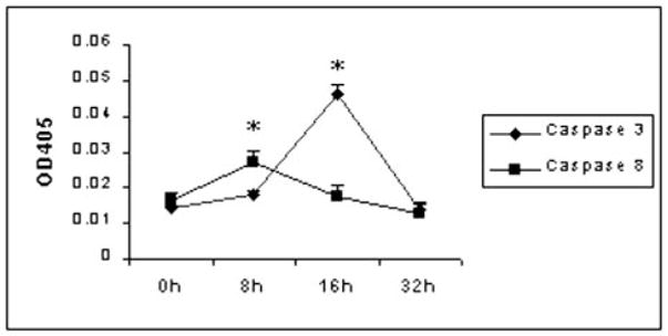
5 × 10⁶ skin-draining lymph node cells from naive C57BL/6 mice were cultured with 60 μg/ml ES products for 0, 8, 16, and 32 h. Cells were harvested at different time points, and caspase-8 or caspase-3 protease activity was examined by a colorimetric protease assay. Data presented are from one of two similar experiments.