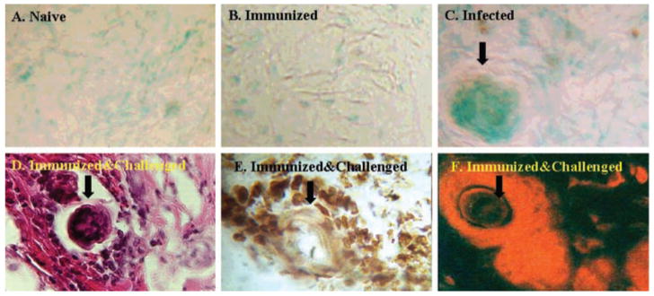
Fig. 3. TUNEL staining for apoptotic cells in the skin.
Skin biopsy samples collected from naive (A), immunized (B), infected naive (C), and immunized and challenged (D–F) mice were processed for cryostat sectioning and stained with biotinylated dUTP (A–C and E), hematoxylin and eosin (D), or PE-labeled anti-CD3 antibody (F). Arrows indicate cut sections of the parasites. The brown-staining cells in C and E are apoptotic cells. Original magnification was ×400. Data presented are from one of three similar experiments using five mice per group.

