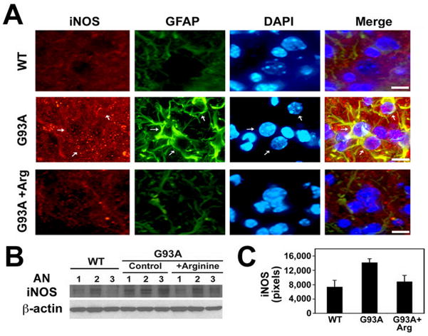
Figure 3. Induction of iNOS level the GFAP-positive cells in the spinal cord of mutant SOD1 (G93A) ALS mice.
A, L-Arginine reduces the immunoreactivity of iNOS in the ventral horn of G93A mice lumbar spinal cord. DAPI was used for counter staining of the nucleus. Arrows indicate iNOS- and GFAP-positive astrocytes. Scale bars: 10 μm. B, Western blot shows that L-arginine supplementation reduces the protein level of iNOS in G93A mice. AN, animal number. The numbers refer to individual animals. C, The densitometric analysis of iNOS protein derived from panel B.

