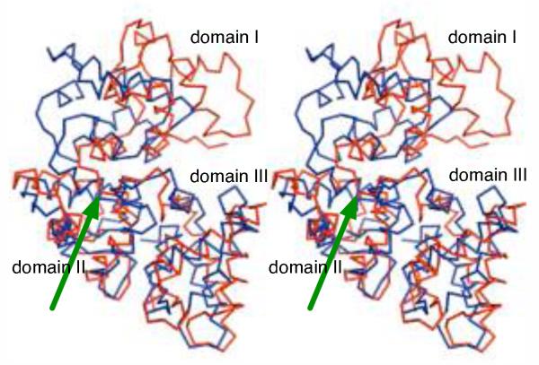
Figure 3.
D. rerio secretagogin and rat calbindin D28K have different quarternary domain arrangement. The domain II and III of D. rerio secretagogin (red) were superposed with the corresponding region of rat calbindin D28K (blue; PDB ID 2f33). In the resulting overlay, the domain I of secretagogin is rotated by almost 180 degrees with respect to the core formed by domains II and III in both proteins. The green arrow points to the point from which structures diverge.

