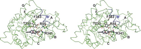
Fig. 6.
Stereoscopic illustration of predicted binding mode of iodoorsellinic acid in CalO2. The carboxyl group is positioned such that a putative pantetheinyl arm would extend from the active site out to helix B' (blue). In this model, C3 of the substrate analogue is approximately 4 Å from the heme iron. Additional helices are labeled as in Figure 4.

