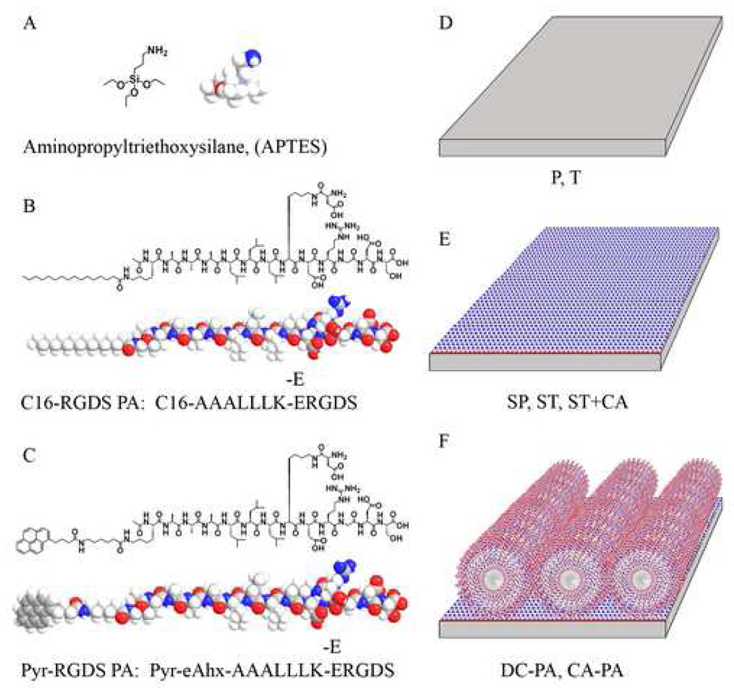
Figure 1.
Chemical structures of APTES (A), the peptide amphiphile (PA) used for AFM, SEM, and biological assays (B), the peptide amphiphile (pyr-PA) used for the fluorimetry assay and AFM (C), and schematics for the various NiTi surfaces obtained in the process to create covalently bound PA nanofibers substrates (D–F).

