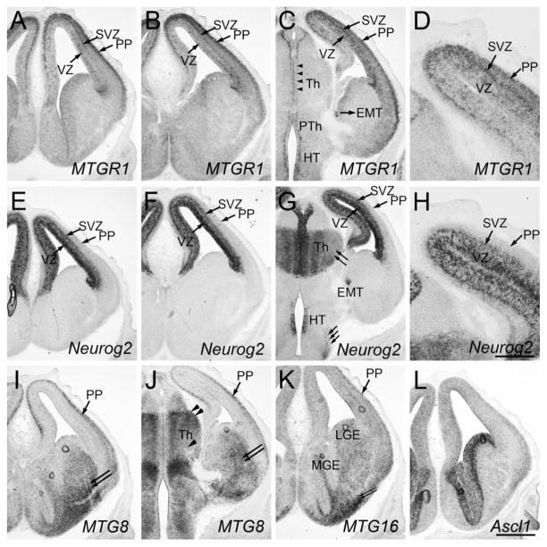
Fig. 3.
Forebrain at E13.5. Frontal sections are shown. A and E, B and F, C and G are adjacent sections. D and H are high-magnification views of C and G, respectively. Midline is to the left. In situ hybridization for MTGR1 (A–D), Neurog2 (E–H), MTG8 (I,J), MTG16 (K), and Ascl1 (L) is shown. PP, preplate; SVZ, subventricular zone; VZ, ventricular zone; MGE, medial ganglionic eminence; LGE, lateral ganglionic eminence; Th, thalamus; EMT, eminentia thalami; HT, hypothalamus. Scale bar = 200 μm for D,H and 500 μm for other panels.

