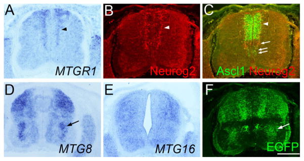
Fig. 9.
Analysis of Ascl1-BAC-EGFP embryos at E11.5. Transverse sections of the lumber spinal cord (A–F) of the same Ascl1-BAC-EGFP embryo at E11.5. Sections in A–F are adjacent to each other and B and C are from the same section. In situ hybridization for MTGR1 (A), MTG8 (D), MTG16 (E), as well as immunostaining for Neurog2 (B), Ascl1/Neurog2 (C), and EGFP (F) is shown. Scale bar = 200 μm.

