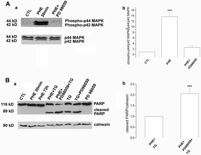
Figure 7. TG-induced apoptosis resistance in DU145 cells is mediated by ERK1/2.
(A)(a) Western blot showing expression of phosphorylated p44/p42 MAPK (ERK1/2) in non-treated cells (CTL), 30 min treatment by 10 µM PHE (PHE 30 min) and simultaneous treatment by 10 µM PHE and 10 µM PD98059 (PHE+PD98059). (b) Expression of phospho-p44 and phospho-p42 MAPK is assessed on total p44/p42 MAPK in DU145 cells. (B)(a) Western blot showing expression of PARP full length fragment (116 kD) and cleaved PARP (89 kD) in the following treatment conditions: non-treated (CTL), 30 min or 72 h treatment by 10 µM PHE (PHE 30 min, PHE 72 h), pre-treatment by PHE alone or in the presence of 10 µM PD98059 followed by 48 h treatment by 10 µM TG (PHE+TG and PHE+PD98059+TG), 48 h treatment by 10 µM TG alone (TG) or preceded by 10 µM PD98059 pre-treatment (PD98059+TG) and 10 µM PD98059 treatment alone. (b) Expression of cleaved PARP is assessed on calnexin used as internal control for PHE+TG and PHE+PD98059+TG treatment conditions. Plots are the average cumulative data (mean ± SEM) of three experiments. Statistical analysis used the t test; ***, p<0.001.

