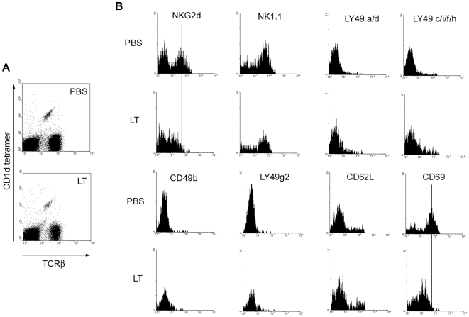
Figure 2. LT alters expression of selected NKT cell surface markers.
C57BL/6 were treated with 100 µg of LT in PBS by the i.v. route or mock-treated with PBS alone. After 96 h, splenocytes were obtained and incubated with FcR-blocking mAb 2.4G2 in the presence of α-GC/CD1d tetramer, anti-TCRβ mAb and mAbs as indicated. Cells were then washed, fixed and analyzed by flow cytometry. A Shows α-GC/CD1d tetramer+/TCRβ+ NKT cells B Histograms show expression of indicated markers after gating on NKT cells. Data are representative of 3 independent experiments.

