Figure 2.
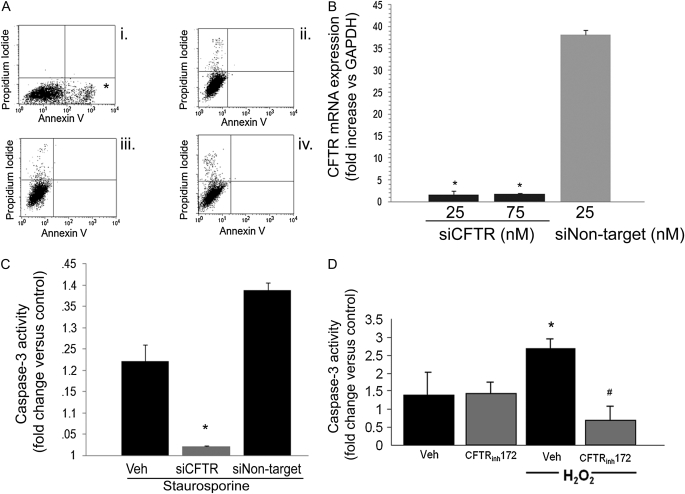
Effect of CFTR inhibitors on stress-induced lung endothelial cell apoptosis. (A) Flow cytometry of mouse lung endothelial cells stained with Annexin V and propidium iodide and treated with (i) staurosporine (0.6 μM, 6 h), (ii) vehicle, and staurosporine with (iii) DPC (200 μM) or (iv) NPPB (200 μM) pretreatments. Note marked increase in apoptotic cells in the right lower quadrant (asterisk) in the staurosporine-treated cells and the profound inhibition of apoptosis in the CFTR-inhibited cells. (B) CFTR mRNA expression (relative to GAPDH) measured in endothelial cells by real-time PCR after transient transfection with siRNA targeting CFTR (siCFTR) at the indicated concentrations or with nontarget siRNA (n = 2; mean ± SD; *P < 0.05 versus nontarget siRNA). (C) Caspase-3 activity in primary human lung microvascular endothelial cells treated with staurosporine (0.6 μM, 6 h) with or without siCFTR (25 nM) or nontarget siRNA (25 nM). Caspase-3 activity was measured in cell lysates as U/μg protein/minute and reported as fold increase versus each respective untreated control (n = 3; mean ± SEM; *P < 0.05 versus nontransfected cells and versus nontarget siRNA-transfected cells). (D) Caspase-3 activity in primary human lung microvascular endothelial cells treated with H2O2 (250 μM, 6 h) in the presence of the CFTR inhibitor CFTRinh-172 (20 μM). Caspase-3 activity was measured in cell lysates and expressed as U/μg protein/min and then reported as fold increase versus control, untreated cells (n = 3; mean ± SEM; *P < 0.05 versus control; #P < 0.05 versus H2O2 treatment).

