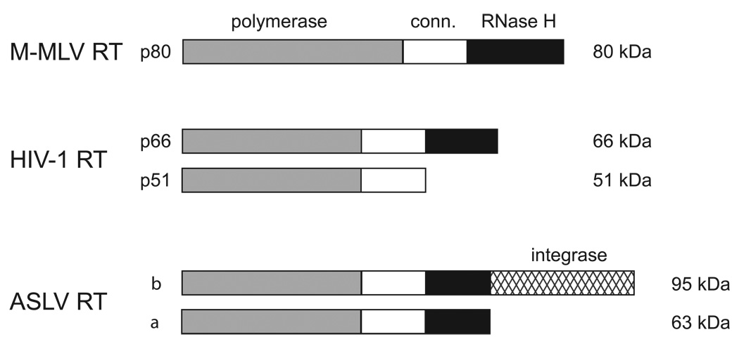
Fig. 1.
Subunit and domain structure of retroviral reverse transcriptases. Reverse transcriptase from M-MLV is a monomer, whereas the HIV-1 and ASLV reverse transcriptases are both heterodimeric. The subunit designations and their sizes (kDa) are indicated along the left and right sides of the figure, respectively. The approximate sizes of the polymerase, connection (conn.) and RNase H domains are shown in gray, white, and black, respectively. The larger β subunit of the ASLV reverse transcriptase also contains the integrase domain depicted by crosshatching.

