Fig. 1.
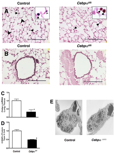
Decreased C/EBPα expression in adult CebpαΔ/Δ mice in type II cells (A) and conducting airway epithelial cells (B). Dams of CebpαΔ/Δ mice were maintained on doxycycline (in chow) from embryonic day 0. CebpαΔ/Δ mice survived postnatally and were treated with doxycycline until 14 days of age. In littermate control mice, C/EBPα was detected in alveolar type II cells (arrowheads) and conducting airways, whereas nuclear staining of respiratory epithelial cells was absent or markedly decreased in CebpαΔ/Δ mice assessed at 7 wk of age. Immunohistochemical staining for C/EBPα was detected in alveolar macrophages (arrows) in both control and CebpαΔ/Δ mice, indicating the specificity of gene deletion in the respiratory epithelium. There were no changes in lung morphology in CebpαΔ/Δ mice under normal conditions. C: decreased expression of Cebpα mRNA in 7-wk-old CebpαΔ/Δ mice lungs. Cebpα mRNA in lung was decreased in CebpαΔ/Δ mice compared with that in control mice analyzed by RT-PCR (n = 3/group). D: decreased C/EBPα protein (by Western blot) in isolated type II cells. The majority of microscopically identified contaminating cells in isolated type II cells was alveolar macrophages in which C/EBPα was detected in CebpαΔ/Δ mice by immunohistochemistry (A). In this system, nearly 70% of Cebpα gene deletion occurs in respiratory epithelial cells from CebpαΔ/Δ mice. Type II cells isolated from 1 male and 1 female mouse were pooled. N = 3 pool/group, *P < 0.01 vs. control. E: electron microscopy was performed on lungs from control and CebpαΔ/Δ mice in room air at 7 wk of age. The ultrastructure of the lung of CebpαΔ/Δ mice, including type II cells, is similar to that of control mice. Shown are representative electron microphotographs of n = 3 mice/group.

