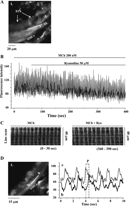
Fig. 4.
The effect of 50 μM ryanodine on Ca2+ signaling induced by 200 nM MCh in airway SMCs. A: a fluorescence image of part of an airway obtained by confocal microscopy under resting conditions. B: changes in intracellular Ca2+ concentration ([Ca2+]i) with respect to time (from a ROI; white square in A) in a SMC in response to 200 nM MCh and 50 μM ryanodine. MCh induced high-frequency Ca2+ oscillations that were unaffected by the addition of ryanodine. Representative data are from ≥10 different slices from 5 mice. C: line-scan plots of the propagated changes in [Ca2+]i (Ca2+ waves) following exposure to MCh (0–30 s) and ryanodine (360–390 s). Line-scans were constructed by sequentially aligning the pixels from the SL shown in A, parallel to the longitudinal axis of the SMC. D: the propagation velocity of the Ca2+ waves was measured by determining the period (P) between sequential changes in Ca2+ in regions “a” and “b” (right) of the same SMCs (left). Ryanodine (50 μM) had no significant effect on the frequency of MCh-induced Ca2+ oscillations, the appearance of the Ca2+ waves, or the propagation velocity of the Ca2+ waves.

