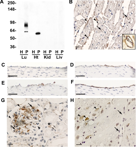
Fig. 2.
J120 immunoreactivity in other normal rat organs. A: Western blot analysis of homogenate and luminal plasma membranes isolated from normal rat lung (Lu), heart (Ht), kidney (Kid), and liver (Liv). Five micrograms of protein were loaded in each lane, and blots were probed with MAb J120. Two bands are seen in lung P, but only one band is apparent in heart and has a slightly faster electrophoretic mobility than the lower-molecular-weight band seen in lung. Molecular weight markers are on the left. B: immunostaining of sections of paraffin-embedded rat heart. Only a few capillaries (arrows) are stained as are fibroblasts and connective tissue elements in the endomysium (arrowheads). Inset shows subendothelial staining in vessels of intermediate size and a lack of staining in the EC. C–F: immunostaining of the aortic arch and the ascending, abdominal, and descending aortas, respectively. Some immunopositive EC are seen in the ascending and abdominal aortas, whereas no staining is seen the EC of the aortic arch. Only in the descending aorta are all cells stained. G: inconsistent staining of capillaries in kidney glomeruli; arrows indicate immunostained capillaries, and arrowheads indicate lack of staining. Some peritubular vessels are also stained (*). H: cerebrum showing J120 immunostaining of neural cells (arrowheads) but not ECs (arrows). Bars in B and G = 10 μm; bars in C–F = 50 μm; bar in H = 20 μm.

