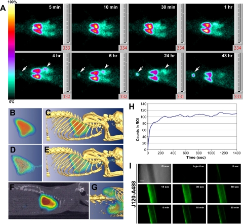
Fig. 3.
In vivo imaging of organ and vessel immunotargeting with labeled MAb J120. A: planar gamma-scintigraphic images acquired at 5, 10, and 30 min, and 1, 4, 6, 24, and 48 h after rats were injected via the tail vein with 125I-J120 (30 μCi at 6 μCi/μg). By 5 min, the antibody has clearly accumulated in the lungs; and at 4 h, new signals in the thyroid (arrow) and stomach (arrowhead) become apparent. No signals were seen in other organs, including heart (*). B–G: tomo graphic scans obtained 30 min after intravenous injection of 30 μCi of 125I-J120 MAb using CT-SPECT. C, E, G: images after fusion of CT isosurface with SPECT. B: volumetric 3-D image of the nuclear signal accumulated in the lung. C: uptake in the lung (from B) correlated with anatomic information from skeleton. D: uptake in the lung (from B) correlated with anatomic information from the thoracic cavity and trachea. E: images from B–D combined. F: sagittal section of CT-SPECT fusion confirms localization of 125I-J120 in thoracic cavity. G: 3-D CT-SPECT fusion showing amplified signal for uptake in kidneys after delineation of lung signal. H: time-course profile (dynamic acquisition) after administration of the 125I-J120 antibody (30-s time frames). Note that the maximum level of uptake is achieved within the first 4 min after injection and remains fairly constant afterwards. I: high-magnification intravital fluorescence microscopy of solitary lung microvessel. Athymic nude mice with implanted rat lung tissue were injected through the tail vein with fluorophore-labeled J120 (J120-A488). The top left image shows a phase-contrast image of a microvessel without other vessels nearby. Fluorescence images were acquired at the postinjection times indicated. Clear endothelial cell-surface binding with J120 is observed, but at no time was extravasation seen. Fading of the J120 signal is due to photobleaching of the fluorophore conjugated to the antibody during the continuous imaging of the microvessel.

