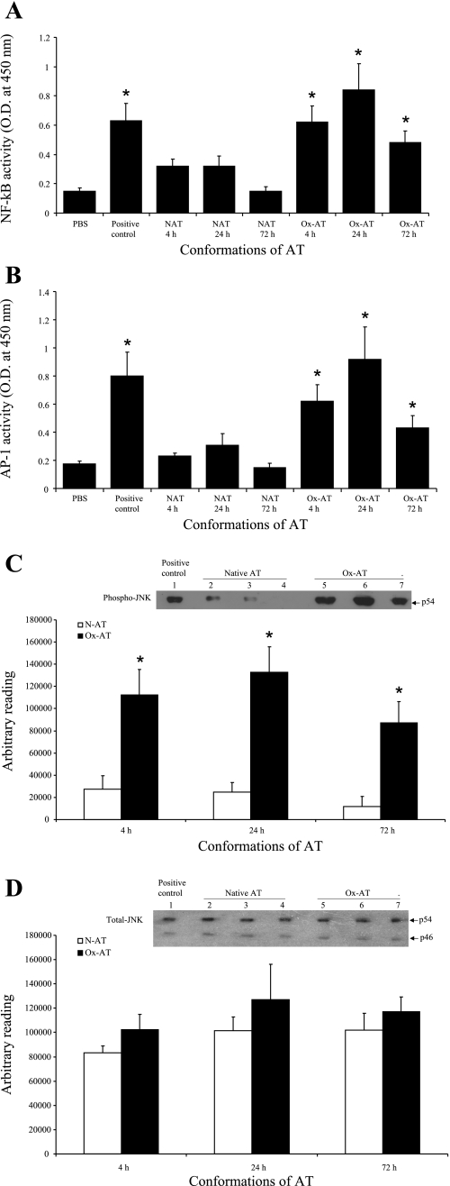
Fig. 9.
A: effect of Ox-AT on induction of NF-κB activity in lung tissue. There was a significant increase in NF-κB activity following intratracheal Ox-AT compared with PBS (4 h, P = 0.028; 24 h, P = 0.029; 72 h, P = 0.037). Intratracheal N-AT did not have any significant effect on NF-κB activity. The positive control was Jurkat cells with high endogenous NF-κB activity (P = 0.032 compared with PBS). Data are means (SE) from 3 separate experiments. *P ≤ 0.05. B: effect of Ox-AT on induction of AP-1 activity in lung tissue. There was a significant increase in AP-1 activity following intratracheal Ox-AT compared with PBS (4 h, P = 0.019; 24 h, P = 0.033; 72 h, P = 0.049). Intratracheal N-AT did not have any significant effect on AP-1 activity. The positive control was K562 cells with high endogenous AP-1 activity (P = 0.022 compared with PBS). Data are means (SE) from 3 separate experiments. *P ≤ 0.05. C: Western blot analysis of homogenized lung tissue for phosphorylated JNK. The p54 JNK isoform was expressed highly in lung homogenates from Ox-AT-treated mice compared with lung homogenates from N-AT-treated mice. Densitometric analysis of each band using ImageJ software demonstrated the level of p54 phospho-JNK was significantly higher and peaking at 24 h in lung homogenates from Ox-AT-treated mice compared with lung homogenates from N-AT-treated mice (4 h, P = 0.031; 24 h, P = 0.012; 72 h, P = 0.023). Lane 1, positive control A431 cell lysate; lane 2, 4 h N-AT; lane 3, 24 h N-AT; lane 4, 72 h N-AT; lane 5, 2 h Ox-AT; lane 6, 24 h Ox-AT; lane 7, 72 h Ox-AT. Gel is representative of 3 independent experiments. Data are means (SE) of 3 independent experiments. *P ≤ 0.05. D: Western blot analysis of homogenized lung tissues for total JNK. Western blot analysis for total JNK shows total (phosphorylated and nonphosphorylated) JNK ranging in size between 46 and 54 kDa. Densitometric analysis of both bands from each sample (expressed cumulatively) using ImageJ software demonstrated total JNK was expressed in comparable levels in lung homogenates from both Ox-AT- and N-AT-treated mice. Densitometric analysis of bands showed there was no significant difference in total JNK in lung homogenates from Ox-AT- and N-AT-treated mice at 4, 24, or 72 h. Lane 1, positive control A431 cell lysate; lane 2, 4 h N-AT; lane 3, 24 h N-AT; lane 4, 72 h N-AT; lane 5, 4 h Ox-AT; lane 6, 24 h Ox-AT; lane 7, 72 h Ox-AT. Gel is representative of 3 independent experiments. Data are means (SE) of 3 independent experiments.

