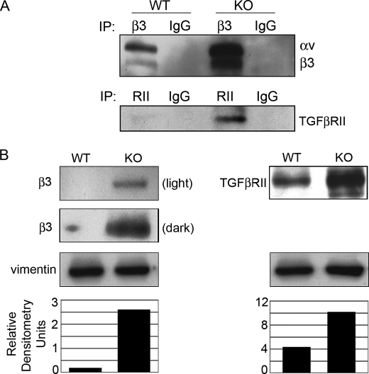
FIGURE 2.
β3 and TGFβRII expression is increased in PAI-1 KO cells. A, cell surface expression. WT and KO cells were biotinylated on the cell surface before lysis and immunoprecipitated for β3 or TGFβRII. Western blots were probed with streptavidin-HRP (SA-HRP) to detect biotinylated proteins. Bands were identified by molecular weight. B, total expression. WT and KO cell lysates were Western blotted for β3 or TGFβRII. Vimentin controls confirm equal loading (densitometry).

