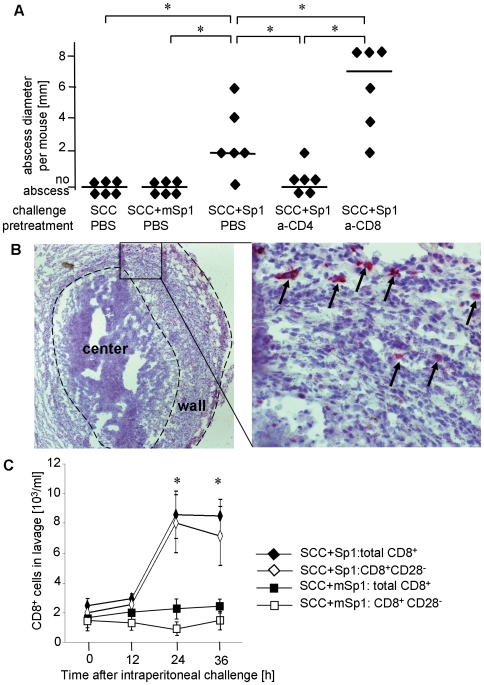
Figure 1. CD8+ T cells modulate abscess formation in response to intraperitoneal Sp1-application.
A. C57BL/6 WT mice received an anti-CD4-mAb, an anti-CD8-mAb, or PBS intravenously 24 h prior to intraperitoneal Sp1-challenge, or challenge with chemically modified Sp1 (mSp1) plus sterile cecal content (SCC) adjuvant, or SCC alone as controls. After 6 days, examination for intraperitoneal abscess formation was performed at autopsy. Each dot represents the abscess diameter per mouse; bars indicate the median abscess size per group; *p<0.05. The experiment was performed three times independently with 6 mice per group, respectively. One representative experiment is shown in the figure. B. Immunohistochemistry of Sp1-induced abscesses was performed with an anti-CD8-mAb. The left panel shows an overview of an abscess. The right panel shows the magnified detail of the abscess wall within the rectangular box of the left panel. Some CD8+ T cells are labeled with black arrows. The figures show one representative result out of three experiments. C. The influx of CD8+ and CD8+CD28− T cells into the peritoneum was determined in C57BL/6 mice challenged with Sp1 or mSp1 plus SCC as a control. The peritoneal lavage was performed at different time intervals (x-axis) after challenge. Cell numbers were determined by cell counting and flow cytometry. Bars indicate standard deviations within a tested group; *p<0.05. The experiment was performed two times with each 4 mice per group and time point. The figure shows one representative result.

