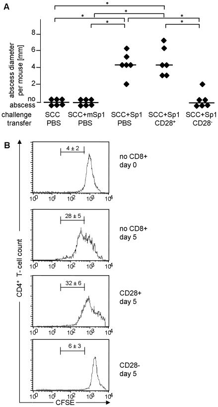
Figure 3. Sp1-induced CD8+CD28− T lymphocytes suppress CD4+ T cell immune responses in vivo and in vitro.
A. Sp1-induced CD8+CD28− T lymphocytes suppress intraperitoneal abscess formation. C57BL/6 mice received intraperitoneal Sp1 challenge (100 µg), and challenge with mSp1 (100 µg), or, SCC adjuvant alone as controls, in the presence or absence of simultaneous intraperitoneal administration of CD8+CD28− or CD8+CD28+ T cells (4×10⁵ cells per mouse). Intraperitoneal abscess formation was examined at autopsy after 6 days. Each dot represents the abscess diameter per mouse; bars indicate the median abscess size per group; *p<0.05. B. Sp1-induced CD8+CD28− T cells suppress CD4+ T cell proliferation in vitro. MLR was performed with DCs from Balb/c mice and CFSE-labeled CD4+ T cells from C57BL/6 mice in the presence or absence of Sp1-induced CD8+CD28− or CD8+CD28+ T cells from C57BL/6 mice. Proliferation of CFSE-labeled CD4+ T cells was evaluated by flow cytometry. The upper histogram shows the result of the incubation of DCs with CFSE-labeled CD4+ T cells for 1 h (control), the lower histograms show the results after 5 days of incubation. The histograms show one representative experiment out of 3 assays performed with cells from three mice each. The percentage of CFSE-positive cells that exhibit proliferation between day 0 and day 5 is given above the marker (±SD; n = 3).

