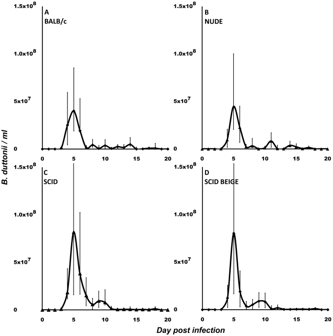
Figure 1. B. duttonii spirochetemia in mice.
BALB/c (A), NUDE (B), SCID (C), and SCID BEIGE (D) mice. B. duttonii established infection in all four mouse strains. Spirochetemia was significantly (p = 0.02) higher in B-cell-deficient mice (C, D). No differences were observed between wild-type and T-cell-deficient mice (A vs. B) or between SCID mice and NK-cell-deficient SCID BEIGE mice (C vs. D). Data are presented as medians with 25th and 75th percentiles. Notice that the scales of the y-axes in this figure differ from those in Figures 2 and 4.

