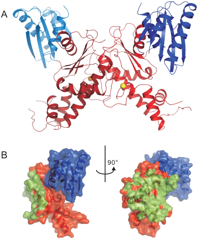
Figure 4. Dimeric GNE kinase domain.
Panel A: Ribbon representation of the dimeric structure of the kinase domain. The N-lobes are shown in blue colors, the C-lobes in red. Panel B: Orthogonal views of the protein surface of one of the monomers. Residues within 4 Å distance from the other monomer are colored green.

