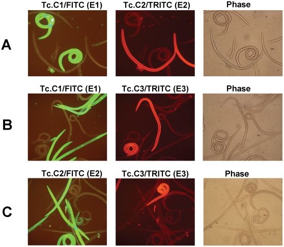
Figure 5. Double-staining immunofluorescence of living T. colubriformis L3 larvae probed with two anti-CarLA scFvs in various epitope group combinations.
Live T. colubriformis exsheathed L3 larvae were incubated with pools of two anti-CarLA scFvs, each recognizing a different epitope and fused to either the E-tag or the c-myc epitopic tag. After washing, bound scFv was stained with both anti-E-tag/FITC mAb and anti-c-myc/TRITC mAb and detected by fluorescence microscopy using filters optimized for FITC (green) or TRITC (red) detection. Within each group (A–C) the same fields were visualized for FITC (left) and TRITC (center) or by phase contrast (right). A. The scFv pool was Tc.C1/E-Tag and Tc.C2/c-myc. B. The scFv pool was Tc.C1/E-Tag and Tc.C3/c-myc. C. The scFv pool was Tc.C2/E-Tag and Tc.C3/c-myc. The epitope group of the anti-CarLA scFvs are indicated in parentheses.

