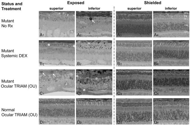
FIGURE 6.
Photomicrographs of plastic-embedded retinal sections from eyes collected 2 weeks after clinical exposure to light. Images were taken approximately 1250 μm from the optic disc along both the superior (A1, A3; B1, B3; C1, C3; D1, D3) and inferior (A2, A4; B2, B4; C2, C4; D2, D4) meridians from the exposed (A1, A2; B1, B2; C1, C2; D1, D2) and shielded (A3, A4; B3, B4; C3, C4; D3, D4) eyes. (A) The untreated mutant (EM109) showed extensive outer retinal degeneration and loss of RPE in the exposed retina, but the shielded retina was normal; (A2, arrow) intraretinal pigmented cell. (B) Systemic DEX (EM110) the photoreceptors and outer retinal layers degenerated in the exposed eye, but the shielded retina was normal. An incomplete, and structurally compromised RPE layer remained (B1, B2, *), and the ONL consisted of one to two incomplete rows of nuclei. (C) Sconj and IVit TRIAM administered to both eyes (OU) of a mutant dog (EM103) before exposure to light did not prevent RPE, photoreceptor, and outer retinal degeneration. Note presence of hypertrophied cells external to the inner nuclear layer (INL) with cytoplasmic inclusions that have cytologic characteristics of macrophages (C1, arrows). The TRIAM-treated shielded, mutant retina remained normal (C3, C4). (D) Intravitreal TRIAM administered to both eyes (OU) of a normal dog (EM100) before exposure to light caused no adverse effects in the exposed or shielded retinas. OS, outer segment layer; IS, inner segment layer; OPL, outer plexiform layer. Scale bar, 20 μm.

