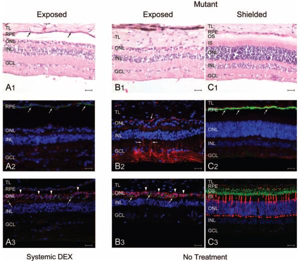
FIGURE 7.
Morphologic and immunohistochemical characterization of the retina from light-exposed (EM108, OS; EM112, OS) and shielded (EM112, OD) T4R RHO mutant dogs; dog EM108 was treated with systemic DEX, and dog EM112 was not treated. All sections from an individual eye were from a series of serial sections taken from the same region. (A1) Marked thinning of the ONL, and loss of photoreceptor inner and outer segments in the DEX-treated, light-exposed mutant retina. Note the preservation of the RPE (arrows). (B1) Similar loss of photoreceptors in the untreated, light-exposed retina. Note, however, the absence of the RPE. (C1) Normal retinal structure and lamination in the shielded eye. (A2) Weak RPE65 labeling (arrows; green) and absence of GFAP labeling (red) in the DEX-treated light-exposed mutant retina. (B2) Strong GFAP labeling in the untreated, light-exposed retina; the absence of RPE65 labeling resulted from RPE loss. (C2) The shielded mutant retina was normal, and RPE labeling was intense and distinct. (A3) The majority of the remaining cell somata in the ONL of the DEX-treated, light-exposed mutant retina were human cone arrestin (hCAR) positive (arrowheads; red); punctate labeling of some cells with the rod opsin antibody is also present (arrows; green). (B3) Similar findings in the untreated, light-exposed retina. (C3) Normal pattern of rod opsin (green) and hCAR (red) labeling in the shielded mutant retina. TL, tapetum lucidum; OS, outer segments; IS, inner segments; INL, inner nuclear layer; GCL, ganglion cell layer. Scale bar, 20 μm.

