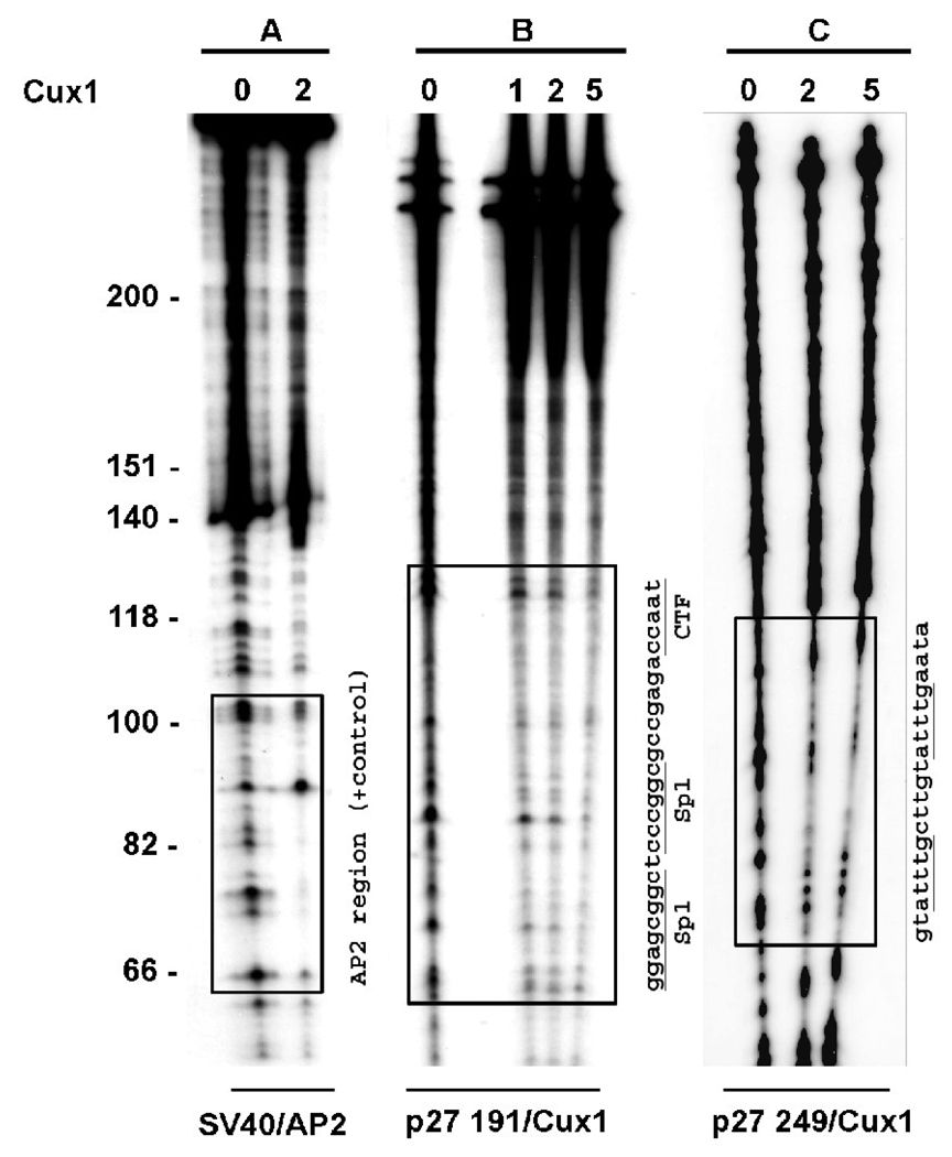
Figure 5. Two separate Cux1 binding sites on the p27kip1 promoter.
DNAse I footprint analysis of the p27kip1 promoter regions bound by Cux1 in vivo. −687 to −496 (panel B) and −1609 to −1360 (panel C) PCR amplification products from ChIP assays were radiolabeled and incubated with purified in vitro synthesized Cux1 protein (amounts shown are in µl) and then digested with DNase1. Regions of DNA protected from DNase1 digestion are indicated by boxed regions. The corresponding sequence for each protected region is shown, with the previously identified Cux1 target sequences underlined. Panel A shows radiolabeled SV40 DNA following incubation with AP2 protein and DNase1 digestion as a positive control. Boxed region shows region of DNA protected from DNase1 digestion.

