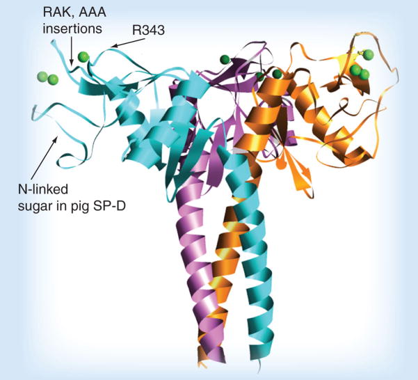
Figure 1. Diagram of the neck and carbohydrate recognition domains of SP-D.
This ribbon diagram was derived from x-ray crystallographic studies of the trimeric binding domain of SP-D in association with the coiled coil neck domain, which is responsible for trimerization. Three saccharide-binding pockets are located on the flat upper surface of the trimer (see spherical single green calcium ion in the pocket). The binding pocket is surrounded on either side by ridges, and substitution of the R343 residue that forms one of the ridges can greatly increase influenza viral-neutralizing activity. Insertions of amino acids (e.g., RAK or AAA, as shown) adjacent to the other ridge also increases antiviral activity to a lesser extent. The location of the N-linked sialylated glycan on pig SP-D is also shown.
SP: Surfactant protein.

