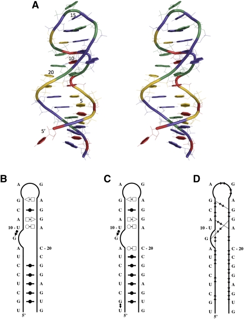
FIGURE 2.
The rat 28S rRNA loop E structure. (A) Stereoview of the crystal structure (PDB code 1Q9A). (Green) Adenosines, (yellow) cytosines, (violet) guanosines, (red) uracils. The thread through the phosphate atoms is shown using a cylinder. Each base ring is filled and highlighted by thick covalent bonds. The H-bonded bases of the characteristic loop E structure, here the G9-U10-A19 base triple, are linked with dotted lines. Note that U1 in this crystal structure is not paired with G27. The image was generated using Pymol. (B) Secondary structure annotated by MC-Annotate. (C) Secondary structure annotated by RNAview. (D) Stacking annotation.

